The revolutionary central nervous system on a chip: how this incredible scientific innovation works
This small chip has the potential to radically change our understanding of the nervous system and offer innovative solutions to medical problems that once seemed insurmountable.

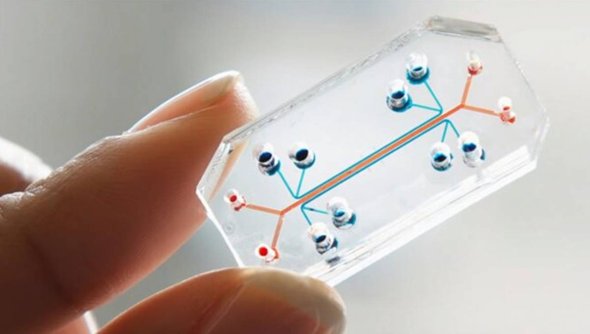
World-renowned researchers have taken a giant step in understanding embryonic development by recreating in the laboratory a miniature and three-dimensional version of the central nervous system, from the brain to the spinal cord. This advancement, led by teams from the Weizmann Institute of Science in Israel and the University of Michigan, utilizes a microfluidic chip capable of simulating the development of the human nervous system at crucial stages of formation.
The development not only opens new doors for studying healthy embryonic growth but also for understanding genetic diseases and tissue damage. This approach represents a new era in developmental biology and organoid engineering.
What is the microfluidic chip and how does it work?
The microfluidic chip is a platform that allows for the replication of the behavior of morphogens, essential molecules in embryonic development. These molecules act as a map that guides stem cells towards their differentiation into specific organs.

Development of the nervous system on a chip: the biology of the future
Unlike traditional methods in Petri dishes, where morphogen concentrations were uniform and limited the formation of complete organs, this chip mimics the natural dispersion of these molecules, creating conditions that simulate the actual development of the human embryo.
How the chip is used:
- Initial preparation: Stem cells are placed on adhesive surfaces of the chip about 4 mm long, simulating the size of a one-month-old embryonic central nervous system.
- Creation of the environment: These surfaces are covered with a gel that emulates the extracellular environment of the embryo, allowing the cells to develop in three dimensions.
- Addition of morphogens: Starting on the third day, morphogens are added at one end of the chip and slowly diffuse through the developing tissue.
- Cell differentiation: Stem cells respond to the varying concentrations of morphogens, organizing into a hollow tube that becomes a replica of the central nervous system, with a cellular arrangement that reflects actual development.
Organoids and their role in scientific advancement
Organoids are miniature versions of organs created in the laboratory. They have revolutionized the study of developmental biology in the last decade, allowing scientists to observe how organs form and respond to diseases.
In this case, the microfluidic chip has enabled the creation of a complete organoid of the central nervous system, from the brain to the spinal cord. This advancement surpasses previous limitations, where it was only possible to create small sections of the nervous system.
Key discoveries with the chip:
- Higher concentrations of morphogens led to cells destined to form the spinal cord.
- Medium concentrations generated the hindbrain and midbrain.
- Lower concentrations resulted in the forebrain, replicating the exact organization of the nervous system in a four-week-old embryo.
Innovation in the creation of cell types
The development of the forebrain was another milestone achieved with this chip. Normally, excitatory and inhibitory neurons, essential for the functioning of the adult brain, must be cultured separately and then combined, a complicated and inefficient process.
Thanks to this technology, researchers were able to generate both cell types in the same tissue. This was achieved by adding morphogens at specific points in the forebrain within the longitudinal tube, replicating natural development.
Result:
- Inhibitory neurons appeared inside the tube.
- Excitatory neurons formed on the outside of the tube, just as occurs in the human embryo.
Impact on the study of diseases
This advancement not only has implications for the study of normal development but also for investigating diseases that affect the nervous system.
Applications of the chip:
- Understanding genetic diseases: The chip allows for the analysis of how specific genetic mutations impact the development of the nervous system.
- Research on neurological disorders: Conditions such as spina bifida or neurodegenerative disorders can be studied at early stages.
- Modeling injuries: Scientists can replicate damage in nervous tissue and test potential treatments more accurately.
The future of embryonic studies
Although the chip does not emulate the early stages of embryonic development, it is capable of advancing stem cells to a state equivalent to that of a four-week-old human embryo. This approach is crucial for studying cellular processes that were previously not fully understood, such as the differentiation of cell populations in the spinal cord.
Moreover, its ability to replicate the three-dimensional formation of tissues opens the door to new research in tissue engineering and regenerative medicine.
The players behind the discovery
This advancement was made possible thanks to the international collaboration of prominent researchers:
- Weizmann Institute of Science: Orly Reiner and Alfredo-Isaac Ponce-Arias from the Department of Molecular Genetics.
- University of Michigan: Jianping Fu and Xufeng Xue, along with their team.
- University of Pennsylvania: Hongjun Song and Guo-Li Ming.
- University of Cambridge: Azim Surani and Frederick CK Wong.
A glimpse into future impact
The microfluidic chip is already helping to answer key questions about the development of the nervous system. Research teams around the world are adopting this technology to expand knowledge about genetic diseases, neurological disorders, and potential treatments.